
Basic Chest CT Interpretation
Nate Beech
Frequent windowing and a consistent, structured search pattern are needed when assessing the various anatomical structures visualized in a standard CT scan of the chest
- Soft tissue window: for evaluation of the organs, soft tissues, and vasculature of the neck, chest, mediastinum, and upper abdomen
- Bone windows: to assess for and osseus lesions or fractures; may also be used to assess for calcifications within the chest (granulomas, lymph nodes, coronary and aortic calcifications)
- Lung window: primary window used when interpreting a chest CT; used to assess the different lobes of the lung for pathology and to assess for free air in upper abdomen
Example of a routine CT chest search pattern:
Lower Neck
- Symmetry within the soft tissues and vasculature,
- Examine for any gross abnormalities within the thyroid gland, cartilaginous structures of the neck, esophagus
- Perfusion deficits (if contrasted study),
- Assess for any subcutaneous air, soft tissue swelling/edema/fluid collections, focal or diffuse lymphadenopathy
- <10mm in the short axis is generally considered normal in the cervical region (numerous exceptions apply based on chain level/clusters/location)
Airways
Examination of the trachea and bronchopulmonary segmental anatomy, assessing for patency, anatomic abnormality, whether trachea is midline.
- Trachea -> carina -> left/right mainstem bronchi -> secondary/lobular bronchi -> smaller bronchi and bronchioles.
Mediastinum
Space containing numerous organs, vessels, nerves, lymphatics that lie within the midline between the chest and lung pleura, extending from posterior of sternum to vertebral column and superiorly from the thoracic inlet to the diaphragm. Divided into anterior, middle, posterior, and superior sections; overall, assess for widening.
- Anterior: contains thymus (if present), lymph nodes, mammary vessels, portions of thyroid gland
- Evaluate for anterior mediastinal mass; differential can include thymoma, thyroid neoplasms, lymphoma, germ cell tumors, vascular abnormalities.
- Middle: contains the pericardium, heart, ascending aorta, pulmonary trunk, right/left pulmonary artery, SVC, mainstem bronchi.
- Assess for malignancy (lymphoma, thyroid mass), lymphadenopathy, thoracic aortic aneurysm, hiatal hernias
- Posterior: thoracic spine, paraspinal nervous system, paravertebral soft tissues, distal azygous venous system.
- Evaluate for malignancy (lung, neurogenic, teratomas, sarcomas, lymphoma), lymphadenopathy, aortic aneurysm.
- Superior: inferior neck muscles, aortic arch, SVC, brachiocephalic veins, trachea, esophagus, thymus (if present), thoracic duct, nervous (phrenic, vagus).
- Assess for malignancy (as above), lymphadenopathy, esophageal pathology (achalasia, hiatal hernia, esophageal neoplasm).
Lymph Nodes
14 total lymph node stations throughout the chest.
- Assess for gross symmetrical or asymmetrical/focal or diffuse lymphadenopathy.
Heart and Great Vessels
Heart, aorta, pulmonary artery and veins, SVC, IVC
- Evaluate heart for cardiomegaly, atrial/ventricular enlargement, pericardial effusion, coronary calcification cavity) of >0.5.
- Cardiomegaly defined as a cardiothoracic ratio (width of cardiac silhouette to thoracic.
- Evaluate great vessels for dilation/enlargement, aneurysm, dissection, pulmonary emboli (seen below), calcification.
- Pulmonary emboli can be evaluated with a CTA PE or V/Q scan, will show filling defect within pulmonary vasculature.
- Acute PE can lead to PEA, right ventricular dysfunction (can be noted by IVC contrast reflux), septal bowing.
- Chronic PE can lead to pulmonary infarction, pulmonary hypertension, cor pulmonale.
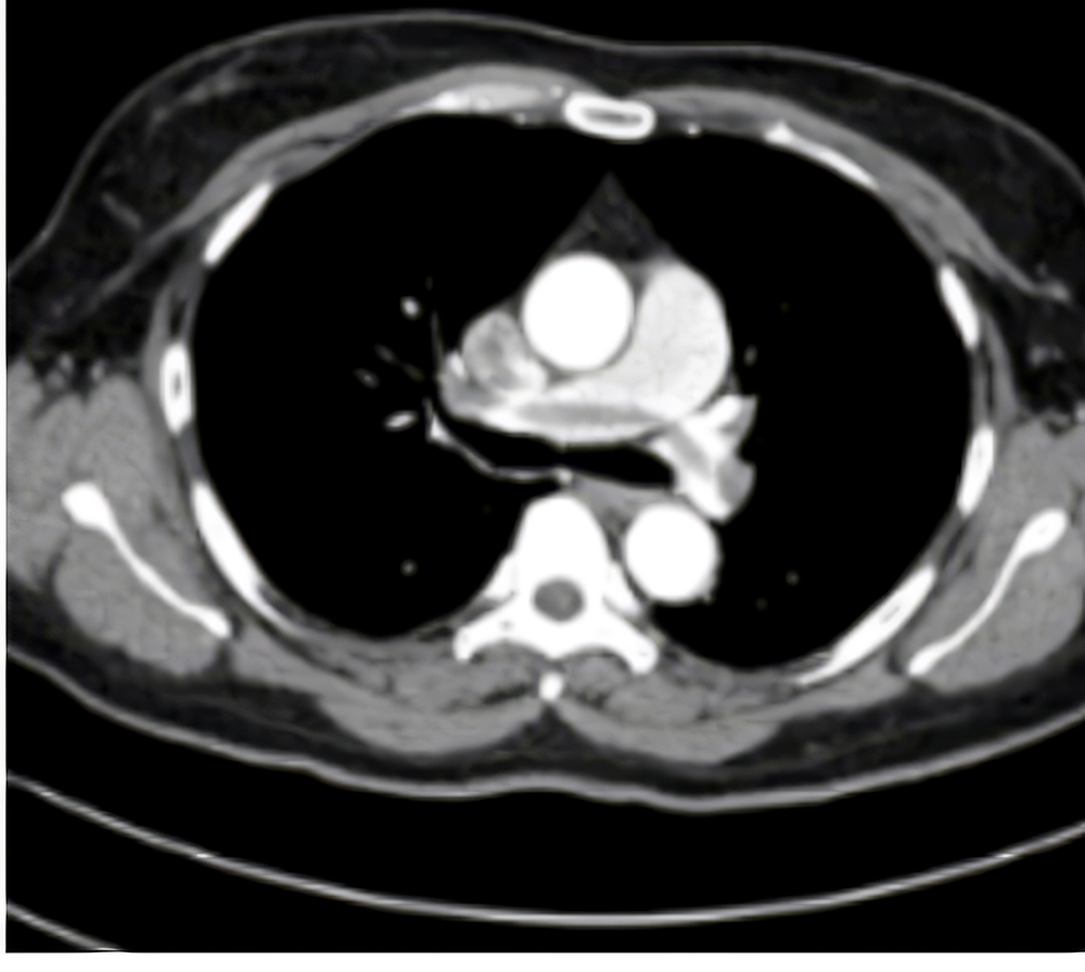
- Thoracic aortic aneurysm: aortic diameter of >4.0 cm in axial diameter, most commonly occur at aortic root or ascending aorta
- Appear as dilation of the lumen with possible thickened walls due to mural thrombus, hematoma/fluid can be seen adjacent if leak or rupture
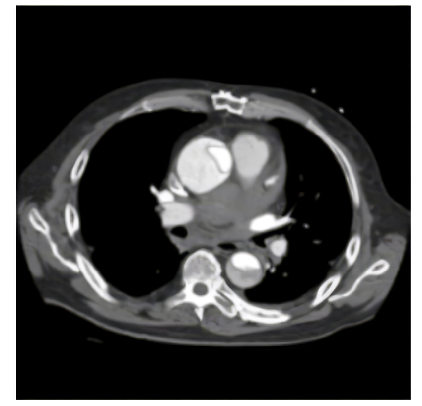
- Thoracic aortic dissection: due to blood entering the medial layer of the aorta through a tear in the intima and tracks longitudinally
- Classified as Standford type A (involves aorta proximal to left subclavian artery) or type B (arises distal to left subclavian artery) [seen below]
- Can use DeBakey system: Type I (ascending and descending aorta), type II (ascending aorta only), type III (descending aorta only, after left subclavian artery)
- Typically see intimal flap, double lumen, aortic dilation, high-density mural hematoma, displacement of atherosclerotic calcification (if present)
- Pulmonary arterial enlargement seen in PE, pulmonary hypertension, connective tissue diseases (Marfan’s, EDS), vasculidities, trauma, infection.
- Defined as dilation of central/proximal pulmonary arteries ≥29 mm in men and ≥27 mm in women.
Lungs
left lung (2 lobes, 1 fissure). Right lung (3 lobes, 2 fissures), and the lingula (some anatomic variability with fissures).
- Evaluate for consolidation, interstitial disease, consolidation, emphysema, atelectasis, nodules, masses, granulomas; identify zone predominance, attenuation, anatomic region (centrilobar, perilymphatic, or random), dominant patter (reticular, nodular, consolidative, cystic).
- Interstitial lung disease: characterized by diffuse cellular infiltrates in periacinar location, causing infiltrative opacification in lung periphery (specific patterns vary depending on etiology).
- Common pathologic causes include granulomatous diseases (sarcoidosis), autoimmune disease (SLE, RA), amyloidosis, idiopathic pulmonary fibrosis.
- Cystic lung disease: groups conditions causing multiple lung cysts, typically seen as thin walled lucencies, with diseases grouped on location (subpleural, intrapleural, intraparenchymal).
- Pathologies in pulmonary fibrosis, pneumocystis pneumonia, Sjögren disease, pulmonary Langerhans cell histiocytosis, amyloidosis, hydatid disease, neurofibromatosis type 1.
- Small airway disease: infectious and non-infectious conditions that affect small airways (peripheral airways usually ≤2 mm) and are associated with air trapping, subsegmental atelectasis, centrilobar nodules, tree-in-bud changes, thickened peripheral airways.
- Common pathologic causes include hypersensitivity pneumonitis, pulmonary sarcoidosis, late/sequela of COVID-19.
- Bronchiectasis: characterized by irreversible dilatation of the bronchial tree and noted by bronchial wall thickening, mucoid impaction, lack of tapering, increased bronchoarterial ratio (diameter of bronchial lumen divided by diameter of accompanying artery), visualization of bronchus within 1 cm of pleural surface, air trapping.
- Typically seen in cystic fibrosis, HIV/AIDS, post-infective (PNA, bronchitis, TB), APBA, asthma, COPD, alpha-1-1antitrypsin deficiency, radiation-induced lung disease, chronic aspiration.
- Consolidation: increased lung attenuation (airspace opacification) sufficient to obscure bronchial walls and blood vessels (non-contrast CT) leaving air bronchograms (seen below).
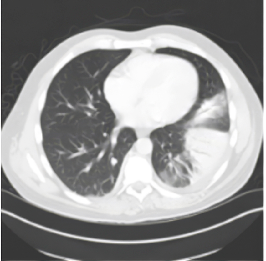
- Generally caused by pulmonary hemorrhage, aspiration pneumonia, bacterial pneumonia, pulmonary edema.
- Air bronchograms: endobronchial air visible against a background of increased lung opacity.
- Atelectasis: small areas of collapsed lung that are not fully expanded characterized by displacement if the interlobar fissures, crowding of pulmonary vessels, crowded air bronchograms, elevation of the diaphragm, displacement of the heart, mediastinum, trachea, hilum.
- Common pathologic causes include mucus plugging, foreign body aspiration, pleural effusion, pneumothorax, radiation fibrosis, granulomatous disease.
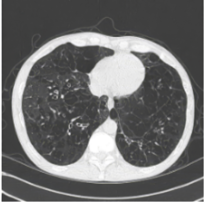
- Emphysema: abnormal enlargement of airspaces distal to the terminal bronchioles with destruction of the alveolar wall (seen below).
- Typically seen in smoking (around 90% of all cases), alpha-1-antitrypsin deficiency.
- Common descriptions of lung findings/pathologies include reticular, nodular, mosaic, ground- glass, tree-in-bud crazy paving, honeycombing, miliary.
- Reticular pattern: excess of linear opacities, resulting from thickening of the interlobular septa or fibrotic changes, either smooth or nodular/irregular thickening.
- Smooth: seen in alveolar proteinosis, pulmonary edema (seen below).
- Nodular/irregular: seen in lymphoma, sarcoidosis, silicosis.
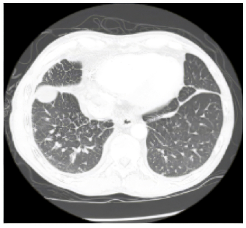
- Nodular pattern: based on distribution of the nodules.
- Perilymphatic: visible in subpleural locations, adjacent to fissures, located in relation to pleural surfaces, interlobular septa, and peribronchovascular interstitium (seen in sarcoid, silicosis, asbestosis, lymphangitic spread of carcinoma).
- Centrilobular: confined to centrilobular region, spare the pleural spaces, typically centered 5-10mm from fissures or pleural surfaces (seen in hypersensitivity pneumonitis, bronchiolitis, infection).
- Random: randomly dispersed without preferences to lung structures or secondary lobule, lack subpleural predominance (seen in TB and fungal infections).
- Tree-in-bud pattern: irregular, nodular branching structure, best visualized at the periphery, representing dilated and impacted centrilobular bronchioles.
- Seen in MAC, TB, bacterial/fungal infection, cystic fibrosis, ABPA, aspiration.
- Ground-glass opacities: represents filling of alveolar spaces with pus, edema hemorrhage, inflammatory cells, tumor with associated thickening of the interstitial or alveolar walls.
- Seen in ARDS, pulmonary edema, PCP, viral pneumonia, organizing pneumonia, lung fibrosis (UIP, NSIP), pulmonary hemorrhage.
- Mosaic pattern: describes density differences between affected and unaffected lung regions, producing a patchy appearance of black and white areas.
- Seen in asthma, PE, bronchiolitis obliterans, hypersensitivity pneumonitis.
- Crazy Paving pattern: combines ground-glass opacities with superimposed septal thickening.
- Seen in alveolar proteinosis, sarcoidosis, organizing pneumonia, infections (PCP, viral, mycoplasma, bacterial), neoplasms, edema.
- Honeycombing: presence of small cystic spaces with irregularly thickened walls composed of fibrous tissue, predominately in the periphery and subpleural lung regions.
- Seen in idiopathic pulmonary fibrosis, RA, scleroderma, asbestosis, end stage sarcoidosis.
- Miliary opacities: innumerable, small 1-2mm hyperdense pulmonary micronodules that are randomly distributed throughout the lung.
- Commonly seen in TB, fungal infections, malignancy.
Pleura
divided into the visceral (covers surface of lung) and parietal (lines inner chest wall) pleura, which enclose the lung and lines the thoracic cavity; typically is not well visualized on CT (thin line along edges of lung) unless pathology disrupts or alters the pleural lining.
- Assess for pneumothorax, pleural effusion, pleural thickening.
- Pneumothorax: gas within the pleural space which separates the parietal and visceral pleura, resulting in lung collapse, usually notable in non-dependent sections of the lung and diagnosed using lung window, which shows area hypodensity between chest wall and collapsed lung (seen below).
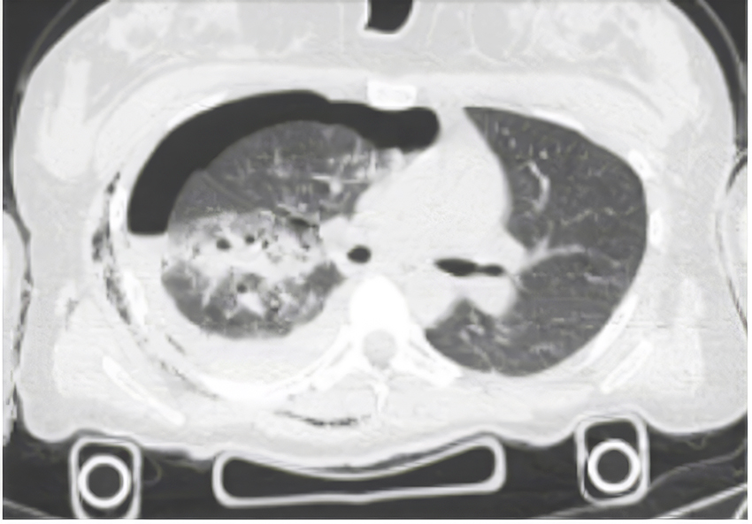
- Typically caused spontaneously, either primary (Marfan’s, EDS), secondary (blebs, asthma, PJP, cystic fibrosis), or iatrogenic or traumatic.
- Pleural effusion: abnormal accumulation of fluid in pleural space, generally collecting dependently and is hypodense (seen below).
- Common causes include heart failure, valvular dysfunction, fluid overload, drowning, ARDS, TRALI.
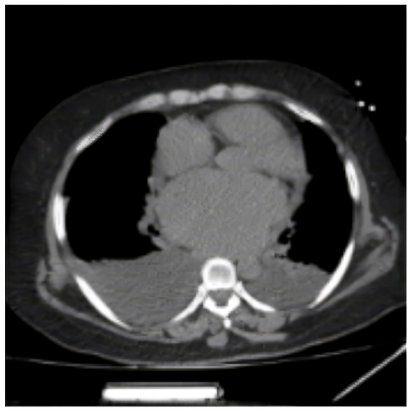
- Pleural thickening: thickening of either the visceral or parietal pleura (either focally or diffusely), resulting in irregular contour.
- Commonly caused by recurrent inflammation, malignancy, mesothelioma, asbestos, talc pleurodesis, recurrent pneumothoracies.
Upper Abdomen
Assess the soft tissues and organs within the upper abdomen including the stomach, spleen, liver, kidneys, pancreas, visualized bowel.
Osseus Structures and Soft Tissues
Spine, neck soft tissues, shoulders (if visualized), clavicles, chest wall structures, abdominal soft tissues.
- Assess for fracture, osseous lesions, masses, trauma, degenerative changes.
Additional Resources for CT Chest Interpretation
- https://radiologyassistant.nl/chest/hrct/basic-interpretation#introduction
- https://radiologyassistant.nl/chest/lung-anatomy/lung-segments
- Felson’s Principles of Chest Roentgenology: a programmed text. Available electronically on Eskind Biomedical Library.
For additional info regarding organ systems/anatomy/disease processes, please see radiopaedia.org reference article.