
Biliary Disease
Sobia Siddiqui
Pearls
- ERCP is not available at VA: requires transfer to VUMC, contact GI to arrange HEMATOLOGY-ONCOLOGY 120.
- Prior cholecystectomy -> CBD normally dilates to 8-10 mm, not necessarily pathological (consider further workup if >10 mm with elevated bilirubin).
- Pneumobilia generally indicates prior biliary sphincterotomy and/or biliary stent.
- CBD dilation classically >6mm, but CBD dilates with age: 70 yo -> 7mm, 80 yo -> 8mm; opiates can also cause biliary dilatation.
Acute Calculous Cholecystitis
- Inflammation of the GB from an obstructing stone in the GB neck or cystic duct.
- Ddx: PUD, pancreatitis, choledocholithiasis, ascending cholangitis, IBD, Fitz-Hugh Curtis.
- Presentation: Severe constant RUQ pain, fever/chills, N/V, + Murphy sign.
- Evaluation: CBC (leukocytosis), CMP (mild AST/ALT ↑), Lipase, Lactate, BCx x2. Tokyo Criteria (2018): need 1x each of local inflammation (pain, Murphy’s sign), systemic inflammation (leukocytosis, fever), imaging (CT or U/S).
- Imaging: RUQ U/S: gallstones + GB wall thickening (>3 mm) or pericholecystic edema.
- If U/S non-diagnostic (no stones or GB inflammation)à HIDA Scan (lack of GB filling).
Management: NPO, IVF, IV Abx until resolved or surgical removal
- Urgent Cholecystectomy (<72H) with EGS.
- If poor surgical candidate: Cholecystostomy with IR; endoscopic drainage options for select patients (i.e. poor surgical candidates also with ascites).
- Complications: gangrenous cholecystitis, perforation, emphysematous cholecystitis, cholecysto- enteric fistula, gallstone ileus.
Acute Acalculous Cholecystitis
Inflammation of the GB without obstructing stone (due to stasis and ischemia)
- Presentation: Seen in critically ill/ICU pts; similar history as above; may present as unexplained fever or RUQ mass (rarely jaundice).
- Ddx: calculous cholecystitis, pancreatitis, hepatic abscess.
- Evaluation: Same as acute calculous cholecystitis.
- Imaging: GB wall thickening, pericholecystic edema, intramural gas, GB distention.
Management: Supportive care, antibiotics, GB drainage
- IVF, correct electrolyte abnormalities, NPO.
- Broad spectrum antibiotic coverage.
- Laparoscopic cholecystectomy is preferred for pts who are not severely ill.
- For severely ill pts: Place CT-guided procedure consult for cholecystostomy placement vs Endoscopic drainage (transpapillary cystic duct stent via ERCP or cholecystoduodenostomy by EUS).
- Consult EGS if necrosis, perforation, or emphysematous changes present.
Biliary Colic
- Transient biliary obstruction typically at the GB neck without GB inflammation (no fever).
- Presentation: Constant (not colicky), intense, dull RUQ pain and N/V for 30 minutes to 6 hours, then resolves, provoked by fatty foods (CCK), absent Murphy’s sign.
- Evaluation: Normal (CBC, LFTs, Lipase, Lactate).
- Imaging: RUQ U/S: cholelithiasis (stones in GB).
- Management: Elective cholecystectomy as outpatient.
Choledocholithiasis
- Obstruction of biliary outflow by CBD stone without inflammation (no fever)
- Impacted cystic duct stone (cholecystitis) with compression of the CBD (Mirizzi syndrome)
- Presentation: RUQ pain (can be painless), N/V and jaundice
- Evaluation: CMP and D-bili (Bili/ALP/ GGT ↑↑↑, AST/ALT mild ↑), CBC (leukocytosis suggests cholangitis), Lipase
- Imaging: RUQ U/S: dilated CBD (>6 mm with GB, >8 mm without GB) à MRCP/EUS vs ERCP (see below)
- MRCP preferred given non-invasive but has lower sensitivity for smaller stones (consider EUS if still have suspicion despite negative MRCP or if patient contraindication to/intolerance of MRI).
Approach to Choledocholithiasis
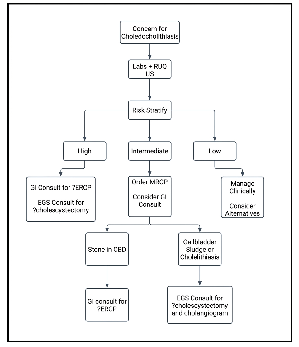
ASGE 2019 Risk Stratification for Choledocholithiasis
| Risk | Clinical and Imaging Features | Management |
|---|---|---|
| High | CBD stone on imaging OR clinical acute cholangitis OR Tbili >4 mg/dL AND dilated CBD (>6 mm with GB, >8 mm without GB) | Proceed directly to ERCP |
| Intermediate | Abnormal liver enzymes OR Age >55 OR Dilated CBD on U/S | EUS, MRCP, or intraoperative cholangiography (IOC) before ERCP |
| Low | No predictors present | Cholecystectomy without IOC |
Management
- NPO & IVF, pain control PRN.
- Procedures as per above algorithm.
- See Acute Cholangitis section If concerned for acute cholangitis.
Acute Cholangitis
- Bacterial infection of biliary tract 2/2 obstruction (typically stones) or prior instrumentation (ERCP).
- Patients with malignant obstruction develop cholangitis less commonly than stone disease, but risk increases significantly after instrumentation (ERCP/stenting).
- Presentation: Charcot triad (RUQ pain, fever, jaundice); Reynolds’ Pentad (+AMS, hypotension).
- Evaluation: CBC, CMP (Dbili, ALP ↑↑↑) Blood Cultures, Lipase, Lactate, CRP, AST/ALT can be ↑↑ as well.
- Imaging: RUQ U/S: dilated CBD (ULN is 6mm), no need for MRCP/EUS prior to ERCP.
- Consider MRCP overnight if ERCP is not being done emergently.
Management
- NPO, IVF, broad-spectrum antibiotics.
- Consult GI for urgent/emergent ERCP (generally within 24-48 hr).
- If ERCP not feasible or fails to establish biliary drainage, can consider EUS-guided biliary drainage, percutaneous transhepatic cholangiography, or surgical decompression.
- Duration of antibiotics: 3-5 days after adequate source control; extend to 2 weeks if Enterococcus or Streptococcus bacteremia (risk of endocarditis).
- Cholecystectomy generally needed within 72 hours of ERCP.